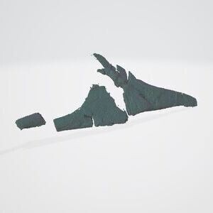
 Media 000394846: Right Jugal
Restricted Download
Type
Mesh
Modality
X-Ray Computed Tomography (CT/microCT)
Media represents biological specimen
Managed by
Media 000394846: Right Jugal
Restricted Download
Type
Mesh
Modality
X-Ray Computed Tomography (CT/microCT)
Media represents biological specimen
Managed by
Login or create an account to download files
GENERAL DETAILS Field Definitions
Media ID
000394846
Media type
Mesh
Object element or part
right jugal
Object represented
Object taxonomy
Navajosphenodon sani
Object organization
Side
--
Orientation
--
Short description
--
Full description
3D model of the right jugal of Navajosphenodon sani (MNA.V.12442, previously MCZ VPRA-9016).
Creator
Tiago Simoes
Date created
February 10, 2020
Date uploaded
November 08, 2021
FILE OBJECT DETAILS Field Definitions
File name
MNA.V.12442_Navajosphenodon_sani_Jugal_Right.stl
File format(s)
STL
File size
56.9 MB
Points
182608
Polygons
364548
Map type
--
UV coordinates
False
Vertex color
False
Bounding box dimensions
7.363, 1.197, 2.520
Centroid coordinates
8.383, 1.637, 14.829
Units of point coordinates
--
IMAGE ACQUISITION AND PROCESSING AT A GLANCE Field Definitions
Number of parent media
1
Number of processing events
2
(2 steps)
Derived directly from
Modality
X-Ray Computed Tomography (CT/microCT)
Device
Zeiss Xradia 620 Versa x-ray microscope (xrm), Center for Nanoscale Systems (Harvard University)
OWNERSHIP AND PERMISSIONS Field Definitions
Data managed by
Data uploaded by
Publication status
Restricted Download
Download reviewer
IP holder
Museum of Northern Arizona
Copyright statement
Creative Commons license
Morphosource use agreement type
Standard
Permits commercial use
Commercial Use Not Permitted
Permits 3D use
3D Printing Limited
Required archival of published derivatives
On MorphoSource
Funding attribution
--
Publisher
--
Cite as
--
Media preview mode
Interactive/Embeddable
Additional usage agreement
None
IDENTIFIERS AND EXTERNAL LINKS Field Definitions
MorphoSource ARK
MorphoSource DOI
External identifier
--
External media URL
--
IMAGE ACQUISITION AND PROCESSING IN DETAIL
STEP 1: IMAGE ACQUISITION
Physical object
mna:v:12442
was imaged to create
undeposited raw media.
IMAGING DETAILS Field Definitions
Modality
X-Ray Computed Tomography (CT/microCT)
Device facility
Center for Nanoscale Systems (Harvard University)
Creator
Tiago Simoes
Event date
--
Software
Dragonfly 4.0
Description
Reconstructed TIFF images of CT scans of MNA.V.12442
Description attachment
None
Reference attachment
None
Exposure time
5
Flux normalization
--
Pixel spacing calibration
--
Shading correction
--
Frame averaging
--
Projections
2022
Voltage
90
Power
--
Amperage
--
Surrounding material
--
X-ray tube type
--
Target type
--
Detector type
Direct (X-Ray photoconductor)
Detector pixels X
--
Detector pixels size X
11.505154
Detector pixels Y
--
Detector pixels size Y
11.505154
Detector configuration
--
Source object distance
--
Source detector distance
--
Target material
--
Rotation number
--
Phase contrast
--
Optical magnification
--
Acquisition type
--
STEP 2: IMAGE PROCESSING
Raw media (undeposited)
was processed
with 1 activity
to create
derived
Media 000392196: Skull And Cervical Vertebrae [CTImageSeries] [CT].
Processing Details Field Definitions
Creator
Tiago Simoes
Event date
September 05, 2019
Software
Dragonfly 4.0
Description
--
Description attachment
None
Processing Activity 1: Reconstruction
Software
Description
Media produced by this event on MorphoSource
STEP 3: IMAGE PROCESSING
Derived
Media 000392196: Skull And Cervical Vertebrae [CTImageSeries] [CT]
was processed
with 1 activity
to create
derived
Media 000394846: Right Jugal [Mesh] [CT] (this media).
Processing Details Field Definitions
Creator
Tiago Simoes
Event date
February 10, 2020
Software
Dragonfly 4.0
Description
--
Description attachment
None
Processing Activity 1: Reconstructed Tomography to Mesh
Software
Description